Understanding the Basics: Defining Electroencephalogram (EEG)
Table of Contents
- Introduction
- Understanding EEG
- Types of EEG
- Procedure of EEG
- Interpretation of EEG results
- Risks and limitations of EEG
- Frequently Asked Questions
Introduction
An Electroencephalogram (EEG) is an essential medical test used by doctors to observe and record the electrical activity of the brain, providing critical insights into various neurological disorders. This process involves placing small, flat metal discs known as EEG electrodes on the scalp that pick up electrical impulses from brain cells and translate them into wavy lines or EEG signals. From diagnosing epilepsy to detecting sleep disorders, an EEG recording plays a pivotal role in clinical neurophysiology by assessing changes in brain waves which can indicate abnormalities.
Taking us back to its inception, Hans Berger, a German psychiatrist developed this groundbreaking technique in the early 20th century. His first human EEG recording in 1924 marked a significant milestone in understanding the functioning of the human brain. Over decades, advancements have been made including flashing lights during EEG tests aiding in diagnosing seizure disorders triggered by light sensitivity or even using ambulatory EEGs allowing patients to engage with their usual activities while monitoring conditions for prolonged duration.
In essence, an EEG test offers a safe procedure that helps detect abnormal brain activity while causing minimal side effects. With ongoing research and technological evolution within this field continues to deepen our knowledge about complex brain disorders and improved patient care methodologies.
Understanding EEG
Electroencephalogram (EEG) is a cornerstone in the field of clinical neurophysiology, providing an invaluable window into the complex electrical activity of the brain. The process involves placing EEG electrodes on the scalp, which capture and convert electrical impulses from brain cells into distinctive wavy lines or EEG signals. These intricate patterns form an ‘EEG recording,’ a real-time visual representation of dynamic brain waves that paints a detailed picture of our neural landscape.
The prowess and precision of this medical test are exceptional in diagnosing neurological disorders such as epilepsy and sleep disorders. A routine EEG can reveal abnormal discharges or show bursts of rhythmic activity that deviate from a normal pattern, signaling a seizure disorder or other neurological conditions. In certain scenarios, flashing lights are introduced during an EEG test to provoke responses, particularly beneficial for diagnosing epilepsy triggered by light sensitivity.
One notable development has been the use of ambulatory EEGs that allow monitoring over prolonged periods while patients engage in usual activities, offering deeper insights into fluctuations in brain activity otherwise masked in short-term evaluations. However brief or extended, every flicker and frequency range captured within an EEG results offers critical data aiding doctors to decipher underlying anomalies.
Despite its sophistication, undergoing an EEG is generally considered a safe procedure with minimal side effects – essentially transforming complex neuronal chatter into actionable medical insights without discomfort to participants. This seemingly simple yet powerful tool continues to revolutionize neurocritical care and our understanding of intricate brain disorders.
Types of EEG
In the dynamic field of clinical neurophysiology, the electroencephalogram (EEG) is a cutting-edge tool that records brain activity. Doctors employ this safe, painless test to chart the electrical impulses produced by neurons in our brains—capturing intricate brain waves and translating them into distinctive wavy lines on an EEG recording. This vital data helps detect abnormal brain activity associated with neurological disorders like epilepsy and sleep disorders. Imagine EEG as a live concert broadcast of your brain’s performance, each band member—a neuron—generating electrical rhythms picked up by ‘microphones’ or EEG electrodes strategically positioned on the scalp.
There are three types of EEG: Routine EEG, Sleep EEG, and Ambulatory EEG. A Routine EEG is akin to a quick soundcheck—it provides doctors with an overview of your brain’s activity but may not capture infrequent anomalies. To record these sporadic glitches, doctors might recommend Sleep or Ambulatory EEGs. A Sleep EEG monitors conditions like sleep disorders during various stages of rest while deep breathing or flashing lights may be used to provoke certain responses. Meanwhile, an Ambulatory EEG allows you to engage in usual activities over extended periods—like having a portable recording studio—that captures how normal daily routines influence electrical activity in your brain.
Each type has its forte in detecting different aspects within frequency ranges and rhythmic activities—from seizure disorders triggered by flashing light sensitivity recorded possibly best via routine tests to abnormalities occurring only during sleep captured through Sleep recording or even fluctuations masked during short evaluations detected effectively via prolonged Ambulatory tests—all critical for accurate diagnoses and effective treatment plans.
| Type of EEG | Description | Use |
|---|---|---|
| Routine EEG | A quick check providing an overview of brain’s activity, may not capture infrequent anomalies. | Best used for detecting seizure disorders triggered by flashing light sensitivity. |
| Sleep EEG | Monitors conditions like sleep disorders during various stages of rest. Deep breathing or flashing lights may be used to provoke certain responses. | Effective for capturing abnormalities that occur only during sleep. |
| Ambulatory EEG | Allows engagement in usual activities over extended periods, capturing how normal daily routines influence electrical activity in the brain. | Best used for detecting fluctuations that might be masked during short evaluations. |
Procedure of EEG
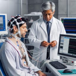
An Electroencephalogram (EEG) is deemed as a cornerstone in clinical neurophysiology, capturing the unseen symphony of electrical impulses that define our brain activity. To prepare for an EEG test, one should be ready for a safe and painless procedure. Hair should be clean and free of any oils or sprays to ensure optimal electrode scalp contact. The actual process involves placing small metal discs called EEG electrodes on the scalp, which record brain waves with precision, transforming them into distinct wavy lines on an EEG recording.
During this medical test, doctors monitor variations in your brain’s performance from normal patterns – think of it as listening to different rhythms in a concert where each neuron contributes to the overall melody. A routine EEG may even expose abnormal discharges or show bursts of rhythmic activity that deviate from the posterior basic rhythm signaling potential neurological disorders like epilepsy or sleep disorders.
Post-procedure too, it’s important to understand that analyzing your ‘brain’s symphony’ can sometimes take time. Your doctor will examine these complex layers of frequency ranges from the recorded data meticulously before discussing results – offering insights into conditions including epilepsy and sleep disorders or even detection of abnormalities such as brain tumours if any.
So while you engage with usual activities post-EEG test, remember this state-of-the-art tool transcends beyond mere diagnostics – it stands testament to modern medicine’s ability to explore our brains’ intricate electrical landscape safely and without side effects – making strides towards advancing neurocritical care every day.
Interpretation of EEG results
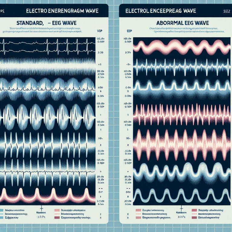
Delving deeper into the realm of electroencephalogram (EEG), it’s fascinating to discover how this medical test, a harmonious symphony of electrical impulses, elegantly records brain activity. Doctors ingeniously decipher these wavy lines or EEG signals to diagnose neurological disorders like epilepsy and sleep disorders. Normal EEG results depict a consistent pattern in frequency range and rhythmic activity—the brain’s melodic concert at equilibrium. However, when seizure disorders hit the high note, abnormal discharges or bursts disrupt this normal pattern. These crescendos in brain waves—captured meticulously on an EEG recording—become flashing lights helping doctors detect abnormal brain activity with precision. Interestingly, even conditions including epilepsy triggered by light sensitivity can be monitored effectively through procedures like routine or ambulatory EEG tests. It’s intriguing how these tests allow patients to engage in usual activities while capturing data for prolonged periods under safe procedural norms—all without causing noticeable side effects! In essence, understanding an EEG is akin to comprehending our brains’ complex orchestra—an interplay of electrical activity creating unique compositions that inform clinical neurophysiology and help enhance neurocritical care methodologies.
| Aspect | Description |
|---|---|
| What is EEG? | Electroencephalogram (EEG) is a medical test that records brain activity as a symphony of electrical impulses. |
| Use of EEG | Doctors use EEG signals to diagnose neurological disorders like epilepsy and sleep disorders. |
| Normal EEG results | Normal EEG results show a consistent pattern in frequency range and rhythmic activity, indicating a brain in equilibrium. |
| Abnormal EEG results | Abnormal discharges or bursts disrupt the normal pattern, often indicating seizure disorders. These abnormalities are captured on an EEG recording. |
| Types of EEG tests | Conditions including epilepsy triggered by light sensitivity can be monitored effectively through procedures like routine or ambulatory EEG tests. |
| Impact on Patients | EEG tests allow patients to engage in usual activities while capturing data for prolonged periods under safe procedural norms, without causing noticeable side effects. |
| Importance of Understanding EEG | Understanding an EEG enhances the comprehension of our brains’ complex orchestra of electrical activity. This understanding informs clinical neurophysiology and helps enhance neurocritical care methodologies. |
| Visualization |
Risks and limitations of EEG
In the realm of clinical neurophysiology, an Electroencephalogram (EEG) is a revolutionary tool that records brain activity with great precision—think of it as a live concert broadcast of your brain’s performance. However, just like any other medical test, EEG also has its limitations and risks despite being generally considered a safe procedure. It falls short in diagnosing certain conditions marked by deep brain abnormalities due to the limitation that EEG electrodes only capture cortical electrical impulses from the scalp surface. Also, while unusual activities are detected effectively during routine or ambulatory EEG tests, problems may arise in interpreting results if individuals have low blood sugar or take specific medications before the test. Indeed, while this non-invasive technology plays an instrumental role in helping detect abnormal brain activity associated with neurological disorders like epilepsy and sleep disorders by capturing every flicker and frequency range within our brains’ symphony, further advancements are needed to overcome these hurdles for improved accuracy and patient care methodologies.